After a fallopian tube cancer diagnosis, your first priorities are to secure the complete pathology record, confirm the cancer’s origin and subtype, and arrange review by a gynecologic oncologist. You will need a staging plan, a clear explanation of surgery versus chemotherapy first, and a practical checklist for comparing treatment teams in Pune.
Key takeaways
- Book a gynecologic oncology consultation, not only a routine gynecology appointment.
- Collect pathology slides, reports, scans, prescriptions and operative notes before your visit.
- Ask where the cancer started and whether pathology confirms fallopian-tube origin.
- Compare upfront surgery with chemotherapy first using stage, operability and surgical goals.
Secure Your Records and Arrange the Right First Appointment
Arrange a gynecologic oncology consultation in Pune promptly, rather than booking only a routine gynecology or benign-laparoscopy appointment. Fallopian-tube epithelial cancer follows treatment principles used for epithelial ovarian and primary peritoneal cancer, so the first specialist should assess cancer staging, operability and the need for chemotherapy.
Take these records to the appointment:
- Biopsy or surgical pathology report, including histologic subtype and immunohistochemistry results
- The laboratory’s information about available tissue slides and paraffin blocks
- CT or MRI reports and the actual image discs or digital files
- CA-125 and other blood-test results
- Prior operation notes, discharge summaries and medication list
- A dated list of symptoms, previous treatments and questions
Ask whether the pathology needs review by a gynecologic pathologist, especially after a small biopsy or an unexpected diagnosis. The review helps distinguish a primary fallopian-tube cancer from cancer that began in the ovary, peritoneum or another site.
The initial visit commonly includes a pelvic examination, contrast CT of the chest, abdomen and pelvis, and blood testing. CA-125 supports assessment and follow-up but cannot diagnose or exclude this cancer alone. Ask about germline testing, including BRCA1 and BRCA2, genetic counseling, and tumour testing for somatic BRCA changes or other treatment-relevant biomarkers.
Verify the specialist’s gynecologic-oncology training, cancer-surgery experience, pathology review process, hospital intensive-care access and multidisciplinary support. That is the practical answer to what to do after fallopian tube cancer diagnosis in Pune.
Confirm the Pathology and Where the Cancer Started
Do not treat the first label as the final answer. Obtain the complete biopsy or surgical pathology report, including histologic subtype and immunohistochemistry (IHC), and request review of the slides and paraffin blocks by a gynecologic pathologist when the diagnosis or origin is uncertain.
Ask the report and review to address:
- Whether the tumour is high-grade serous carcinoma or another histology, because subtype affects treatment and prognosis.
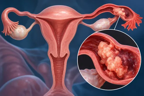
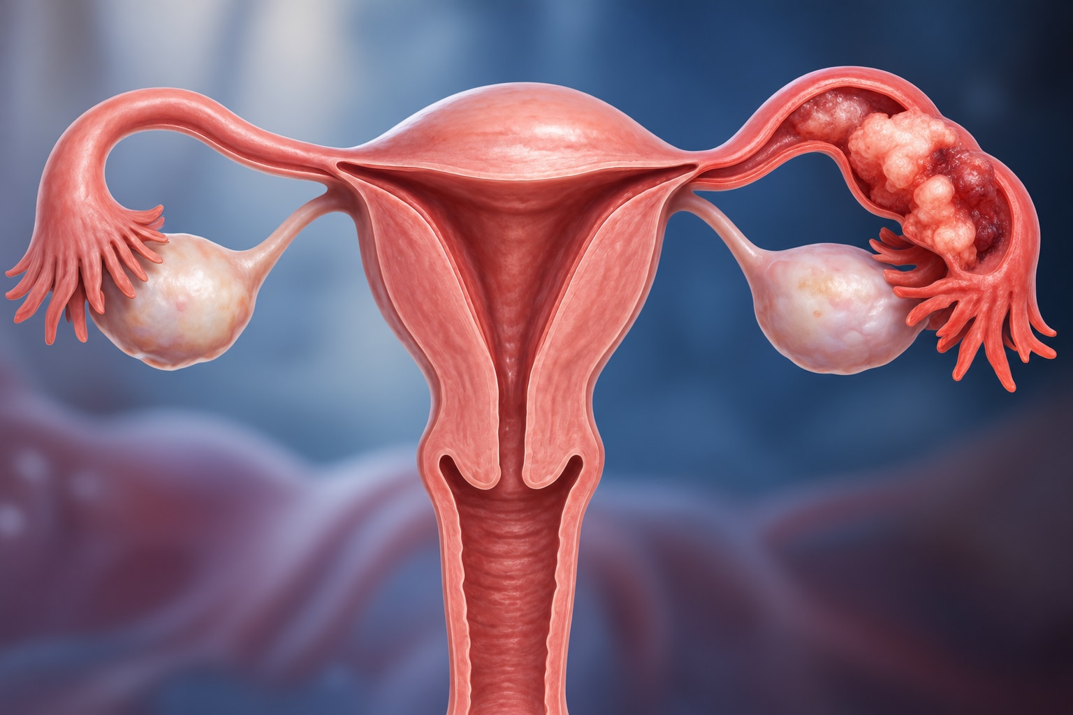
- Whether the fallopian tube contains the dominant tumour, mucosal involvement or serous tubal intraepithelial carcinoma (STIC), a possible precursor lesion.
- Whether the ovaries, peritoneal surfaces or another organ show a larger or more convincing primary tumour.
Doctors combine these findings with pelvic examination and contrast CT of the chest, abdomen and pelvis. A mass centred in the tube with characteristic tubal involvement supports primary fallopian-tube cancer; a dominant ovarian mass or widespread peritoneal disease without a convincing tubal precursor may indicate ovarian or primary peritoneal cancer.
The distinction is not always absolute, and epithelial cancers from these sites are staged and treated using closely related principles.
CA-125 can support the assessment and provide a baseline for follow-up, but it cannot diagnose or exclude tube cancer alone. Arrange review with a gynecologic oncologist rather than relying only on a general gynecologist. Searches such as “fallopian tube cancer specialist Aundh” identify a location, not cancer-surgery training, so verify pathology review and gynecologic-oncology experience.
Understand the Staging Tests and What They Mean
FIGO stage is estimated with examination and scans, but confirmed most accurately from operative findings, biopsies and lymph-node pathology. A gynecologic oncology consultation in Pune should decide whether complete macroscopic resection appears feasible and whether your health permits major cytoreductive surgery.
| Test | What it establishes | Treatment implication |
|---|---|---|
| Pelvic examination | Detects pelvic masses, fixation or visible disease | Supports surgical planning but cannot exclude abdominal spread |
| Contrast CT of chest, abdomen and pelvis | Maps peritoneal deposits, lymph nodes, liver or lung involvement, ascites and disease distribution | Extensive or difficult-to-remove disease supports chemotherapy before surgery |
| MRI pelvis | Gives better detail about pelvic organs and local invasion | Helps define whether pelvic disease can be removed safely |
| PET-CT | Clarifies selected indeterminate lesions or suspected distant spread | Used when CT or MRI leaves an important staging question |
| CA-125 and blood tests | CA-125 provides a baseline for monitoring; blood counts, kidney and liver tests assess treatment fitness | A raised CA-125 does not prove cancer or establish FIGO stage |
Ask the team to state the likely stage: I is limited to the tubes or ovaries, II involves other pelvic structures, III includes abdominal peritoneum or retroperitoneal nodes, and IV includes distant spread.
If imaging suggests complete upfront removal is unlikely, or major surgery is unsafe, platinum-taxane chemotherapy followed by interval debulking surgery is commonly considered. Ask why surgery first is recommended—or why chemotherapy first offers the safer route.
Compare Surgery, Chemotherapy First and Surgical Routes
The decisive question is whether the team can remove all visible cancer safely. Tumour spread, residual disease risk, your medical fitness and the surgeon’s experience—not the surgical instrument alone—determine the plan.
| Option | What it means | When it applies |
|---|---|---|
| Open surgery | A larger abdominal incision gives access to the pelvis, omentum and upper abdomen for staging and cytoreduction. | Bulky disease, extensive upper-abdominal or peritoneal spread, suspected lymph-node involvement, or a high chance of needing complete debulking. |
| Laparoscopic or robotic surgery | Small incisions and a camera allow staging or removal of selected disease while preserving cancer-surgery principles. | Limited disease, no expected need for extensive resection, and a surgeon who can avoid tumour rupture and remove all visible cancer. |
| Chemotherapy first | Platinum-taxane chemotherapy precedes interval debulking surgery, followed by further chemotherapy. | Imaging suggests complete upfront cytoreduction is unlikely, or heart, lung or other health problems make major surgery unsafe. |
Surgical staging can include washings, biopsies of suspicious peritoneal areas, removal of the uterus, both tubes and ovaries, omentectomy and selected lymph-node assessment. Leaving visible residual disease is linked with poorer outcomes, so a smaller-incision approach is the wrong choice if it prevents complete resection or safe access.
Ask why primary surgery is recommended or rejected. Seek a second opinion with the actual scans and pathology when extensive cytoreduction or robotic surgery for advanced disease is proposed, rather than relying on a brief summary.
Use Your Pune Consultation to Test the Treatment Plan
Before you consent, ask the team to explain the plan in writing: FIGO stage, histologic subtype, whether complete macroscopic resection is feasible, and why surgery or chemotherapy comes first.
1. “Can you remove all visible disease?” Ask what imaging shows, which organs might require removal, the planned route—open, laparoscopic or robotic—and what would make the surgeon stop rather than leave unsafe residual disease.
2. “Why this sequence?” Clarify the expected number of platinum-taxane chemotherapy cycles, whether interval debulking surgery is planned, and how your medical conditions change the risk. Chemotherapy first does not mean the cancer is untreatable.
3. “Which tests affect later treatment?” Request germline genetic counseling and testing, including BRCA1 and BRCA2, plus tumor testing for somatic BRCA changes and other relevant biomarkers. Ask how results affect relatives and maintenance treatment.
4. “How will complications be handled?” Identify the hospital’s intensive-care access, blood-bank arrangements, interventional radiology, pathology support and plan for bowel, urinary or vascular complications.
5. “Who is leading the case?” A local specialist should demonstrate gynecologic-oncology training, experience with cytoreductive surgery, pathology review and multidisciplinary tumour-board care—not merely offer a convenient location. Search terms such as fallopian tube cancer doctor baner and fallopian tube cancer consultation pashan identify clinics, not competence.
Dr kunaal Shinde - Best Gynecologist - 08048037552 belongs in your comparison only after you verify these details directly. Seek a second opinion with imaging and pathology review before extensive surgery, neoadjuvant chemotherapy, fertility decisions or robotic surgery for advanced disease.
Related service
Gynec Cancer Treatment Best Fallopian Tube Cancer Specialist in Baner, Pashan, Aundh, Balewadi,Pune Best Fallopian Tube Cancer Specialist in Pune | Advanced Robotic & Laparoscopic Cancer Surgery | Dr. Kunaal K. View service → |
Frequently asked questions
Which specialist should you see after a fallopian tube cancer diagnosis in Pune?
Arrange a gynecologic oncology consultation. The specialist should assess staging, operability and whether chemotherapy is needed before surgery.
What records should you take to your first appointment?
Take pathology reports and slides or tissue blocks, scan images and reports, blood-test results, operative notes, discharge papers and a current medicine list.
How is fallopian tube cancer confirmed and distinguished from ovarian or primary peritoneal cancer?
Ask the pathology team to review the diagnosis and identify where the cancer started. Fallopian-tube epithelial cancer follows treatment principles used for epithelial ovarian and primary peritoneal cancer.
What staging tests might be discussed after diagnosis?
Your specialist may review cross-sectional imaging, blood tests and pathology findings to assess disease spread, stage and whether complete surgery is feasible.
How do doctors compare surgery first with chemotherapy before surgery?
The decision depends on the stage, disease distribution, your fitness for surgery and the likelihood of removing all visible cancer. Ask the specialist to explain the treatment goal and alternatives.
Keywords