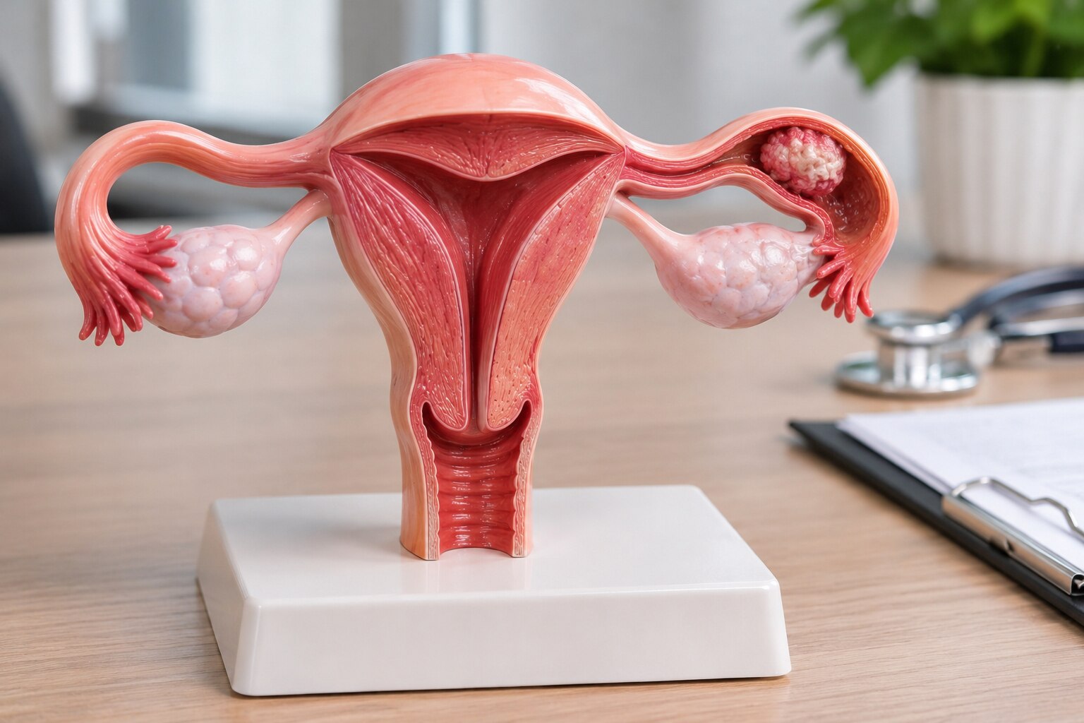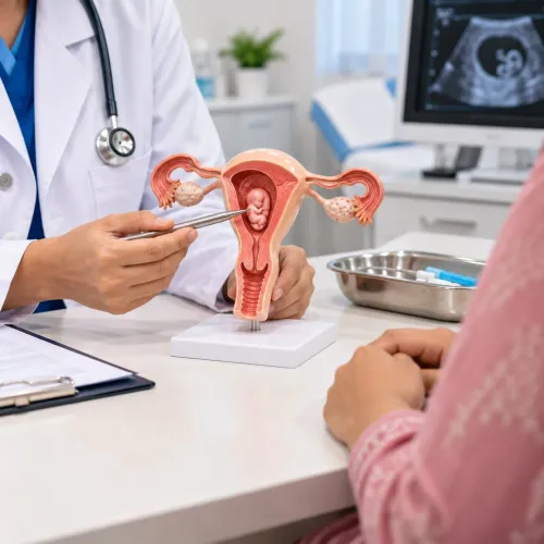
A fallopian-tube mass, an elevated CA-125 result and a confirmed cancer diagnosis lead to different decisions, so no single operation or chemotherapy plan fits every patient. By the end, you will know which test findings establish cancer, how FIGO stage and pathology alter treatment, and what to ask when comparing surgery and systemic care in Pune.
Key takeaways
- Confirm the cancer’s starting site using examination, imaging and tissue findings.
- Use the pathology report and FIGO stage to guide treatment decisions.
- Match chemotherapy and surgery to stage, overall health and surgical fitness.
- Discuss fertility-sparing or minimally invasive surgery with a gynecologic oncologist.
How doctors confirm where the cancer started
Doctors confirm the starting site by combining examination, imaging and tissue findings; no single scan or blood test can do it. Fallopian-tube cancer follows the epithelial ovarian and primary peritoneal cancer pathway because these cancers behave similarly, yet doctors still investigate whether the tube, ovary, uterus or another organ was first involved.
1. A pelvic examination checks for tenderness, fullness or a mass. Transvaginal ultrasound then assesses both tubes, ovaries, uterus and pelvic fluid.
2. CT of the chest, abdomen and pelvis maps enlarged nodes, peritoneal deposits, fluid and distant spread. MRI helps clarify a pelvic mass when ultrasound cannot show its origin clearly.
3. CA-125 provides a clue, not a diagnosis. Endometriosis, fibroids, menstruation, pelvic infection and other cancers can raise it, while a normal result cannot exclude cancer.
4. Hysteroscopy is useful when bleeding or imaging suggests disease inside the uterine cavity, especially suspected endometrial cancer. Laparoscopy allows direct inspection of the abdomen; doctors can take biopsies, collect peritoneal fluid or washings, and assess whether disease began in the tube or spread from elsewhere.
5. A pathologist experienced in gynecologic malignancy establishes the diagnosis from tissue. The review distinguishes a primary tubal mass from ovarian cancer, endometrial cancer extending outward and metastatic disease.
That distinction can change the operation, chemotherapy sequence and genetic or tumour testing. It explains how fallopian tube cancer care changes with diagnosis in Pune: treatment follows histology, spread and fitness, not the anatomical label alone.
What the pathology report and FIGO stage change
A biopsy can identify cancer, but the final treatment plan depends on what the tissue shows and where the disease is found. Record the histologic type, grade, depth of invasion, lymphovascular invasion, and whether serous tubal intraepithelial carcinoma (STIC) is present. STIC is a precursor lesion strongly associated with high-grade serous carcinoma.
1. High-grade serous, low-grade serous, endometrioid, clear-cell and mucinous carcinomas do not behave identically or respond to treatment in the same way. Ask for review by a pathologist experienced in gynecologic malignancy when the subtype or grade is uncertain.
2. FIGO stage I means cancer is confined to one or both tubes. Stage II means spread to other pelvic organs; stage III means peritoneal spread outside the pelvis and/or retroperitoneal lymph-node involvement; stage IV includes distant disease, such as malignant pleural effusion or organ metastasis.
3. Symptoms, scans, CA-125 and a needle or fluid sample provide clues before treatment; they do not always establish the final stage. Surgical assessment adds peritoneal washings, biopsies, omentum examination or removal, and selected pelvic and para-aortic lymph-node assessment.
4. Final pathology can upgrade an apparently early cancer when washings contain malignant cells, microscopic invasion appears, or nodes, omentum or biopsies reveal spread. That result changes whether care resembles early-stage surgery or combined surgery and systemic treatment.
For fallopian tube cancer care planning in Pune, review the report with gynecologic oncology, pathology and medical oncology teams. Base decisions on histology, FIGO stage, spread pattern and fitness—not the anatomical label alone.
How stage and fitness determine chemotherapy and surgery
For apparently stage I disease, surgery usually confirms the stage as well as removes cancer. The operation is tailored to histology, grade, visible spread and fertility goals.
| Stage | Surgical approach | Systemic treatment |
|---|---|---|
| Stage I | Remove the affected tube and ovary; assess the opposite side; collect peritoneal washings; inspect the abdomen and biopsy suspicious areas; remove the omentum; assess selected pelvic and para-aortic lymph nodes. | Chemotherapy depends on histology, grade, invasion and final staging. |
| Stage II–IV | Plan cytoreductive surgery with the goal of no visible residual tumour when this is safe. | Platinum-based chemotherapy, commonly carboplatin with paclitaxel, before or after surgery. |
For stage II, III or IV disease, fallopian-tube cancer usually follows the epithelial ovarian-cancer pathway. If scans or diagnostic laparoscopy predict incomplete or unsafe tumour removal, chemotherapy first followed by interval surgery can reduce surgical risk.
Assessing fallopian tube cancer treatment suitability in Pune requires more than reviewing the stage: kidney function, blood counts, nutrition, other illnesses and recovery capacity affect both chemotherapy and the timing of surgery.
Offer germline testing, including BRCA1 and BRCA2, alongside tumour testing for homologous-recombination findings. After a platinum response, selected patients may receive maintenance with a PARP inhibitor and/or bevacizumab. The decision depends on biomarkers, prior treatment, blood pressure, kidney function, blood counts and other toxicity risks; maintenance does not replace effective surgery or chemotherapy.
When fertility-sparing or minimally invasive surgery is suitable
Fertility-sparing surgery is reserved for unilateral, carefully staged early disease with favourable pathology and a strong pregnancy goal. It is not the default for fallopian-tube cancer. High-grade or invasive cancer, spread beyond one tube, bilateral involvement, or a hereditary risk that calls for broader risk reduction can make preservation unsafe.
| Option | Main benefit | What must be true |
|---|---|---|
| Fertility-sparing surgery | Preserves the uterus and a functioning ovary when possible | Disease is unilateral, early, favourable in pathology, and complete staging remains possible |
| Laparoscopic or robotic surgery | Smaller incisions and often faster recovery | The team can stage and remove the specimen intact without rupture or tumour spillage |
| Open laparotomy with definitive surgery | Wider access for complete tumour removal | Large or adherent masses, upper-abdominal or disseminated disease, or visible tumour at multiple sites |
Complete staging involves peritoneal washings, inspection of the abdomen, directed biopsies, omentectomy and selected pelvic and para-aortic lymph-node assessment. A minimally invasive label proves nothing if the operation cannot establish the stage or remove all visible disease.
When definitive treatment is safer, surgery may include removal of the uterus and both fallopian tubes and ovaries. That approach causes infertility and, after ovary removal, premature menopause; discuss egg or embryo preservation before treatment when time and cancer safety permit.
Open surgery is often safer when disease is extensive or organs are stuck together. The decisive questions are tumour distribution, visibility, intact specimen extraction and whether the surgeon can achieve no gross residual disease—not the technology name.
How to plan specialist care in Pune after the report arrives
Bring the entire record to the first specialist visit, because a missing slide, image or operative note can delay the treatment decision. Carry:
- Pathology slides or tissue blocks and the complete pathology report
- CA-125 results in date order, not only the latest value
- Ultrasound, CT and MRI images with their reports
- Medication list, allergies and major medical conditions
- Details of prior operations, including discharge summaries
Ask whether the cancer is primary tubal, ovarian, endometrial extending outward or metastatic. Ask which FIGO stage is established and which remains suspected, and whether a pathologist experienced in gynecologic malignancy has reviewed the tissue.
| Primary cytoreduction | Neoadjuvant chemotherapy | When medical optimisation comes first |
|---|---|---|
| Immediate tumour-reduction surgery, followed by chemotherapy | Chemotherapy before interval cytoreductive surgery | Significant anaemia, poor nutrition, uncontrolled illness or limited fitness for either route |
| Consider when complete visible tumour removal appears safely achievable | Consider when imaging or laparoscopy predicts incomplete or unsafe surgery | Improve fitness, then reassess the treatment sequence |
Ask how the team will monitor carboplatin-paclitaxel timing, blood counts, kidney function, recovery, treatment response and delays. Request multidisciplinary review involving gynecologic oncology, medical oncology, radiology and pathology, with genetics and fertility counselling when relevant.
When comparing a fallopian tube cancer specialist in Pune, ask about formal gynecologic-oncology training, staging and cytoreduction experience, no-gross-residual-disease rates in comparable advanced cases, complication management and access to the wider team.
Dr kunaal Shinde - Best Gynecologist - 08048037552 is one local option for discussing open, laparoscopic or robotic suitability; pathology, stage, spread and fitness should decide the approach, not the technology label.
Related service
Gynec Cancer Treatment Best Fallopian Tube Cancer Specialist in Baner, Pashan, Aundh, Balewadi,Pune Best Fallopian Tube Cancer Specialist in Pune | Advanced Robotic & Laparoscopic Cancer Surgery | Dr. Kunaal K. View service → |
Frequently asked questions
How do doctors confirm where fallopian-tube cancer started?
Doctors combine pelvic examination, imaging and tissue findings to assess whether the cancer began in the fallopian tube, ovary, uterus or another organ.
How do the pathology report and FIGO stage change treatment?
The pathology report identifies the cancer type and features, while the FIGO stage shows how far it has spread; together they guide surgery, chemotherapy and follow-up planning.
How do stage and fitness determine chemotherapy and surgery?
Doctors weigh the cancer’s stage, tumour distribution, overall health, organ function and ability to tolerate anaesthesia before recommending chemotherapy, surgery or both.
When is fertility-sparing or minimally invasive surgery suitable?
Suitability depends on cancer stage, tumour location, spread, fertility goals and whether removing less tissue can still provide safe cancer treatment.
How should you plan specialist care in Pune after receiving the report?
Take the pathology report, imaging discs and written reports, operation notes and medication list to a gynecologic oncology consultation for coordinated treatment planning.
Keywords