An ovarian cyst does not cause fallopian tube cancer or prove that cancer is present, but the two conditions can appear together and produce similar symptoms. By comparing anatomy, risk factors, ultrasound findings, and follow-up tests, you can understand when routine observation is reasonable and when you need prompt specialist assessment.
Key takeaways
- An ovarian cyst does not cause fallopian tube cancer.
- Persistent, complex, or enlarging masses need medical assessment.
- Ultrasound checks a cyst’s size, shape, contents, and blood flow.
- A suspicious mass may require specialist review and tissue diagnosis.
Does an ovarian cyst cause fallopian tube cancer?
No—an ovarian cyst does not cause fallopian tube cancer or, by itself, increase your risk of developing it.
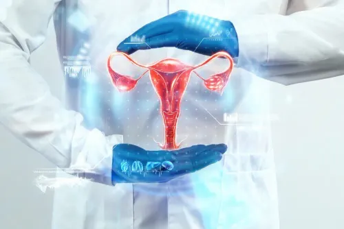
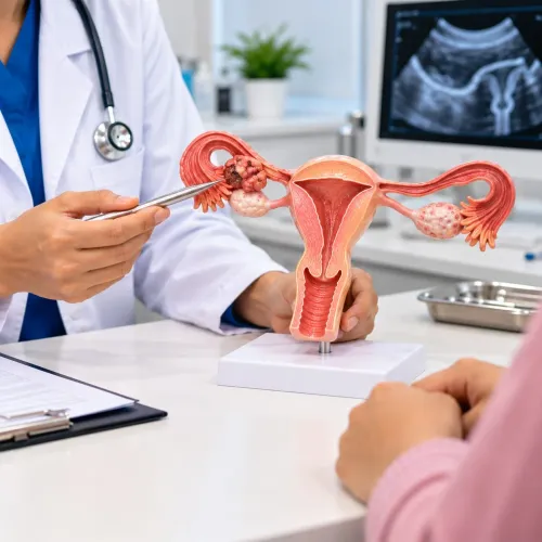
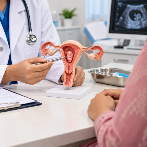
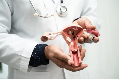
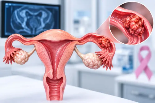
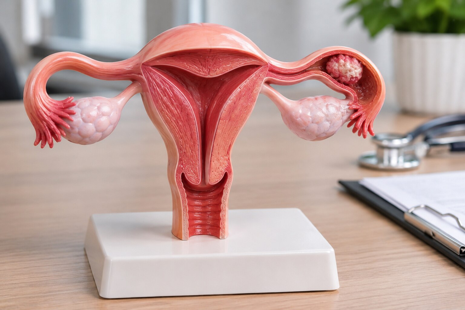
1. Identify where the abnormality starts. An ovarian cyst is a fluid-filled sac within or on an ovary. An ovarian tumour is a solid or mixed growth arising from the ovary. A fallopian-tube mass begins in the tube, while a para-tubal, or paratubal, cyst sits beside the tube or ovary rather than inside either organ.
2. Consider the common explanation first. Functional cysts are common before menopause and often disappear without treatment. A cyst can exist at the same time as unrelated fallopian tube cancer, but finding a cyst does not show that it caused cancer or predicts that cancer.
3. Understand why doctors examine the whole area. Primary fallopian tube cancer is uncommon, and some high-grade serous cancers once labelled ovarian cancer are now linked to serous tubal intraepithelial carcinoma, or STIC, near the fimbrial end of the tube. Doctors therefore assess both ovaries, both tubes, and the peritoneum when a mass looks suspicious.
4. Treat the ultrasound finding as a starting point, not a diagnosis. A simple cyst often supports observation, while a persistent, enlarging, solid, irregular, or complex lesion needs proper evaluation. Symptoms such as pain, bloating, or pressure overlap between conditions.
For anyone researching fallopian tube cancer and ovarian cysts in Pune, persistence and unusual scan features matter more than the word “cyst” alone.
Which symptoms and personal factors make a cyst more concerning?
The overlap between fallopian tube cancer symptoms and ovarian cysts makes symptoms an unreliable diagnosis. Both conditions can cause pelvic or abdominal pain, bloating, increasing abdominal size, pressure, urinary frequency, bowel changes, early fullness, abnormal vaginal bleeding, or watery or blood-stained vaginal discharge.
Persistent or progressive symptoms need examination even when a scan shows a cyst.
| Finding | Why it matters | What to do |
|---|---|---|
| Rapidly increasing abdominal swelling or persistent pain | A cyst may be enlarging, or another pelvic process may be present | Arrange prompt medical assessment |
| Postmenopausal bleeding, a new complex mass, or unusual discharge | These findings deserve more than routine cyst monitoring | Seek earlier gynecological review |
| Sudden severe one-sided pain with nausea or vomiting | This pattern suggests ovarian torsion | Obtain urgent medical assessment |
| Abrupt pain after a cyst ruptures | Rupture can cause internal bleeding | Go for urgent assessment, especially with dizziness or weakness |
Age and menopause status change the level of concern. A new, enlarging, persistent, or complex mass after menopause deserves structured evaluation rather than watchful waiting alone.
Personal history also matters. Tell the clinician about breast, ovarian, fallopian tube, or related cancers in close relatives, a BRCA1 or BRCA2 variant, or any previous cancer diagnosis. These factors can justify earlier specialist involvement even when symptoms are mild.
What does an ultrasound reveal about a cyst or tube-area mass?
A transvaginal ultrasound usually comes first because it examines both ovaries and the fallopian-tube region. A simple cyst is thin-walled and fluid-filled, with no solid areas, papillary projections, thick septations, or internal blood flow.
| Scan appearance | What it may indicate | Usual next step |
|---|---|---|
| Simple cyst with clear fluid | Lower concern | Observation or repeat ultrasound when advised |
| Complex mass with solid parts, irregular walls, papillary projections, thick septations, bilateral lesions, ascites, or marked Doppler blood flow | Higher concern | Further assessment and specialist review |
| Large, distorted, or tube-adjacent lesion that cannot be clearly classified | Indeterminate finding | Follow-up imaging, often MRI |
These features raise concern but do not prove cancer. A mass can remain unclear when its size or position prevents the ultrasound specialist from seeing its origin. Follow-up imaging shows whether it resolves, persists, or enlarges; MRI can better characterise an indeterminate mass.
CT is used more often to assess suspected spread or plan cancer treatment than to investigate every uncomplicated cyst.
If you are searching ovarian cysts cancer concerns in Pune, remember that a normal-looking scan cannot override worsening pain, bloating, unusual discharge, bleeding, or a high-risk history. Sudden severe one-sided pain with nausea or vomiting needs urgent assessment because torsion or rupture can occur independently of cancer.
How is fallopian tube cancer evaluation carried out in Pune?
A structured fallopian tube cancer evaluation in Pune starts with risk and symptom assessment, not a tumour-marker result. The clinician reviews pain, bloating, bleeding or unusual discharge, menopause status, family history of breast, ovarian, fallopian tube or related cancers, and any previous cancer diagnosis before performing a pelvic examination.
1. Transvaginal ultrasound examines both ovaries and the tube region. Doppler assesses blood flow. The report should identify whether the mass is simple or complex, persistent or enlarging, and whether it has solid areas, papillary projections, thick septations, irregular walls, ascites, or bilateral disease.
2. Blood tests can include CA-125 when examination and imaging make it clinically appropriate. CA-125 is not a stand-alone cancer test: menstruation, endometriosis, fibroids, pelvic infection, liver disease, and other benign conditions can raise it, while some cancers do not. Interpret it with imaging and menopausal status.
3. Do not use routine CA-125 or transvaginal ultrasound screening if you have average risk and no symptoms. False-positive results can lead to unnecessary scans, procedures, and surgery.
4. If the mass looks suspicious, seek gynecologic-oncology referral before surgery. Specialist planning can change staging, specimen removal, and protection against tumour spillage. Dr kunaal Shinde - Best Gynecologist - 08048037552 can provide one local point for coordinated assessment and referral when needed.
Related service
Gynec Cancer Treatment Best Fallopian Tube Cancer Specialist in Baner, Pashan, Aundh, Balewadi,Pune Best Fallopian Tube Cancer Specialist in Pune | Advanced Robotic & Laparoscopic Cancer Surgery | Dr. Kunaal K. View service → |
What happens when a mass remains suspicious?
A small, simple cyst in a premenopausal woman can be monitored with repeat ultrasound when her symptoms, examination, and scan all support observation. The plan changes when the mass persists, enlarges, causes symptoms, looks complex, or appears after menopause.
Before choosing treatment, ask:
- Is the lesion ovarian, tubal, or paratubal?
- Does it contain solid tissue, papillary projections, thick septations, or internal blood flow?
- Are there ascites, lesions on both sides, or signs of spread?
- Was CA-125 interpreted alongside menopausal status, examination, and imaging? Endometriosis, menstruation, fibroids, pelvic infection, and liver disease can raise it, while cancer can occur with a normal result.
- Should a gynecologic oncologist plan the operation?
A planned cyst procedure may be appropriate for a persistent but benign-appearing mass. Laparoscopy or robotic surgery can mean smaller incisions and faster recovery in selected cases, but minimally invasive access does not prove that the mass is benign.
If cancer is suspected, safe removal and complete staging take priority. Tumour size, spread, and the risk of rupture can change the surgical route.
Searches for fallopian tube cancer and ovarian cysts in Pune, or for fallopian tube cancer evaluation in Pune, should lead to risk-based specialist assessment rather than a diagnosis based on the word “cyst.” Women with ovarian cysts can also develop fallopian tube cancer, but the cyst itself neither proves nor predicts that cancer.
Frequently asked questions
Does an ovarian cyst cause fallopian tube cancer?
No. An ovarian cyst does not cause fallopian tube cancer or, by itself, increase your risk of developing it. A cyst and a fallopian tube cancer are different conditions, although imaging may require further assessment when findings overlap.
Which symptoms and personal factors make a cyst more concerning?
Persistent pelvic or abdominal pain, increasing abdominal size, unusual vaginal bleeding, unexplained weight loss, early fullness, or urinary and bowel pressure deserve medical review. Concern also rises with a complex or enlarging mass, postmenopausal status, a strong family history of ovarian or related cancers, or a known hereditary cancer risk.
What does an ultrasound reveal about a cyst or tube-area mass?
A pelvic ultrasound assesses the mass’s size, location, solid and fluid-filled parts, wall thickness, internal projections, septations, blood flow, and free fluid. Transvaginal ultrasound often provides the clearest view of the ovaries, fallopian tubes, and surrounding pelvis.
How is fallopian tube cancer evaluation carried out in Pune?
Evaluation usually starts with a gynecological examination and pelvic ultrasound. Depending on the findings, a specialist may request repeat imaging, MRI or CT, blood tests such as CA-125, and review of personal and family history. These tests do not replace tissue diagnosis when cancer remains a concern.
What happens when a mass remains suspicious?
A gynecologic oncology team reviews the imaging and test results, then discusses observation, additional imaging, laparoscopy, or surgery. If tissue is removed, a pathologist examines it to determine whether the mass is benign, borderline, or malignant and to guide treatment.
Keywords